Imagine the vibrant world of a hawk soaring through the skies or a cat stalking through tall grass. Their keen eyesight is crucial for survival. But what happens when a life-threatening injury or disease targets one of their most vital senses? Whether it’s a majestic owl injured by a stray branch or a loyal dog suffering from a painful eye infection, these creatures rely on the skill and compassion of veterinary professionals to restore their quality of life. Enucleation, the surgical removal of an eye from the orbit, is sometimes the only solution to save these animals from pain and further complications. While it might seem drastic, this surgery can offer a second chance at life, whether for a wild predator, prey or a beloved pet when there is irreversible damage to the eye.
Last June, we encountered a striking example of this when a fragile, infant male white-tailed deer was brought to our center in a dire state. The fawn had first been spotted in this condition a few days earlier, struggling and swimming up a lake before collapsing on the shore, visibly injured. Despite his obvious distress, people left him there alone for several days, hoping his mother would come get him. Unfortunately, this delay only worsened his condition as he laid there, cold, wet, alone and suffering from his injuries. It’s important to note that if anyone encounters an animal in such a state, it’s crucial to contact us or another wildlife rehabilitator immediately, rather than waiting, because prompt intervention can be life-saving.
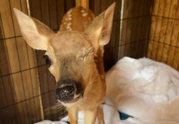
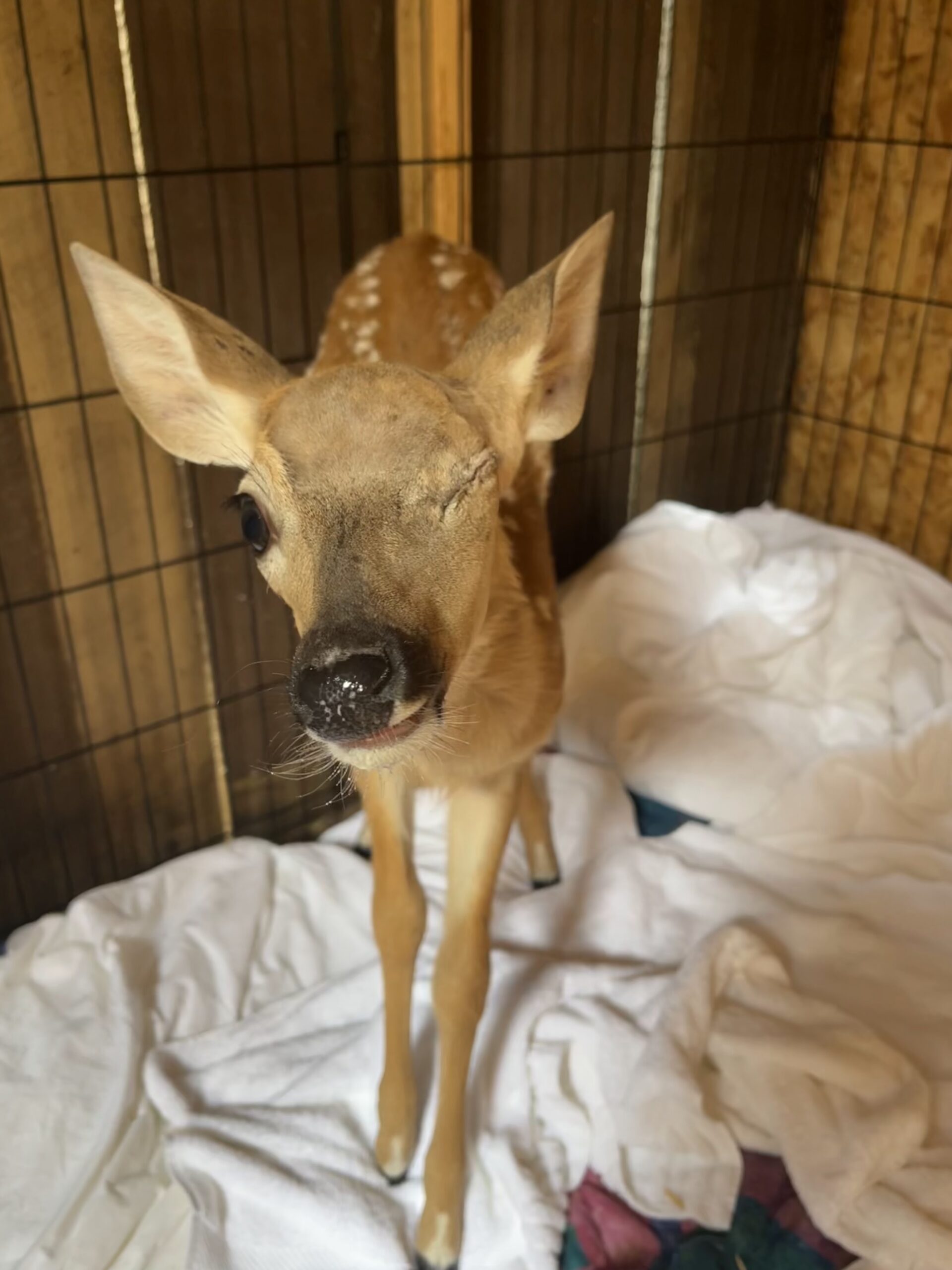
After those agonizing days, the fawn was finally brought to our facility, where he could receive the urgent care he desperately needed. Severely dehydrated and barely responsive, his tiny body bore the marks of a brutal encounter, likely with a predator or possibly even a dog attack. He had multiple puncture wounds scattered all across his head, legs, and body, some already starting to get infested with maggots. However, his most serious injuries were a severe head trauma, due to which he could not even stand on his own, and his left eye, which also had a serious trauma injury.
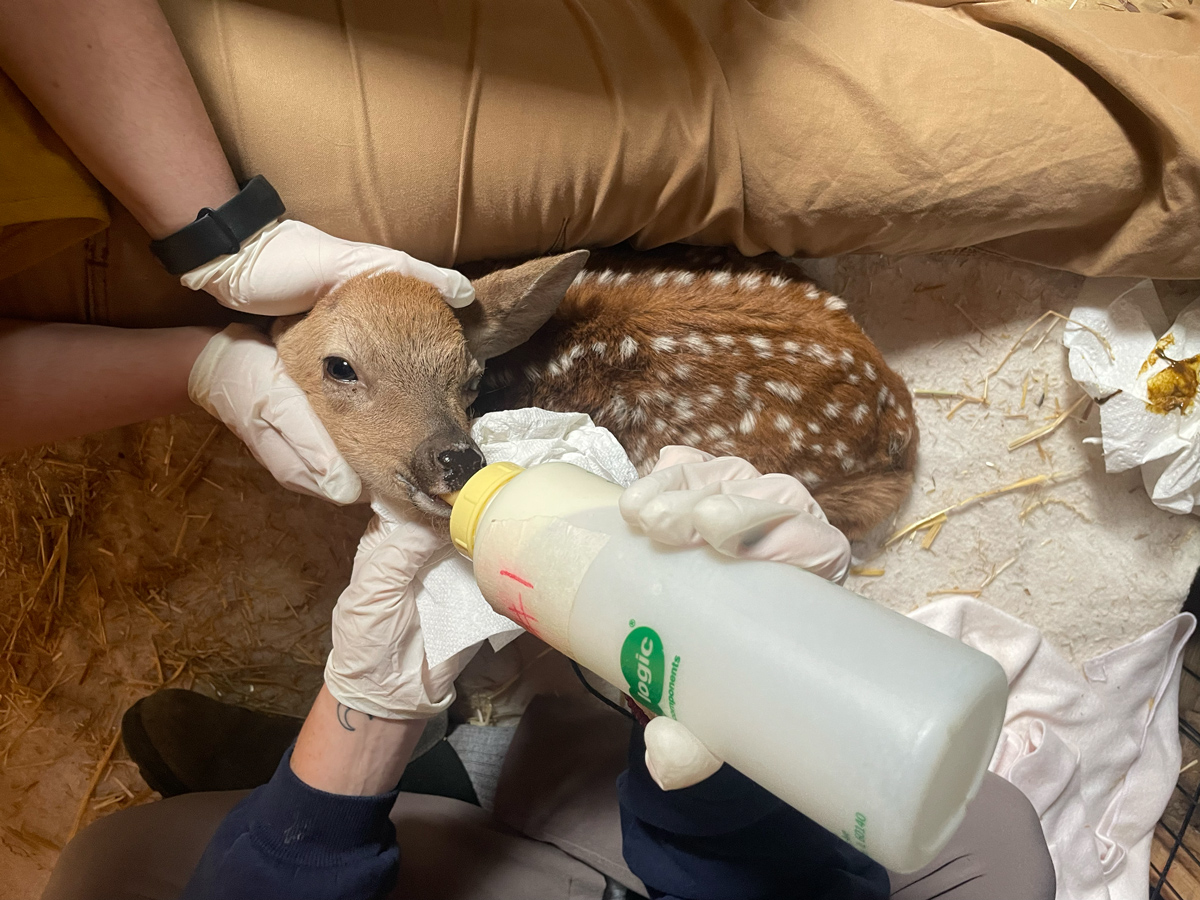
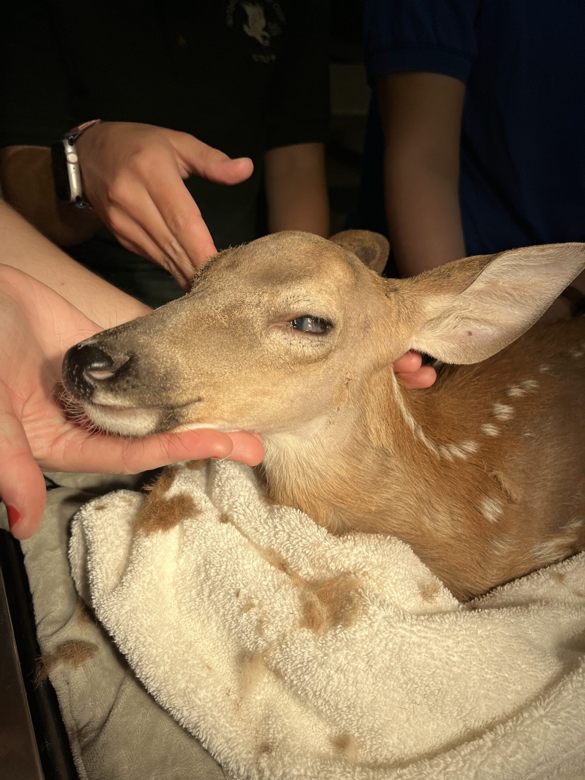
Our first priority was to stabilize the fawn. We administered antibiotics, pain meds, anti-parasitic, and a medicated eye ointment. Over the next month or so, we focused solely on his recovery, helping him build his strength. He slowly started improving and was also finally able to stand on his own! However, despite our efforts, it became clear that his injured eye required more than just medication. In the beginning, we knew that the road to his recovery would be long and uncertain, but now that the fawn had a few weeks to stabilize and had regained some of his strength, we could consider more invasive treatments for his eye because there was a big possibility that the damaged eye could turn into even greater complications in the future such as glaucoma or other eye diseases that would be a source of immense pain.
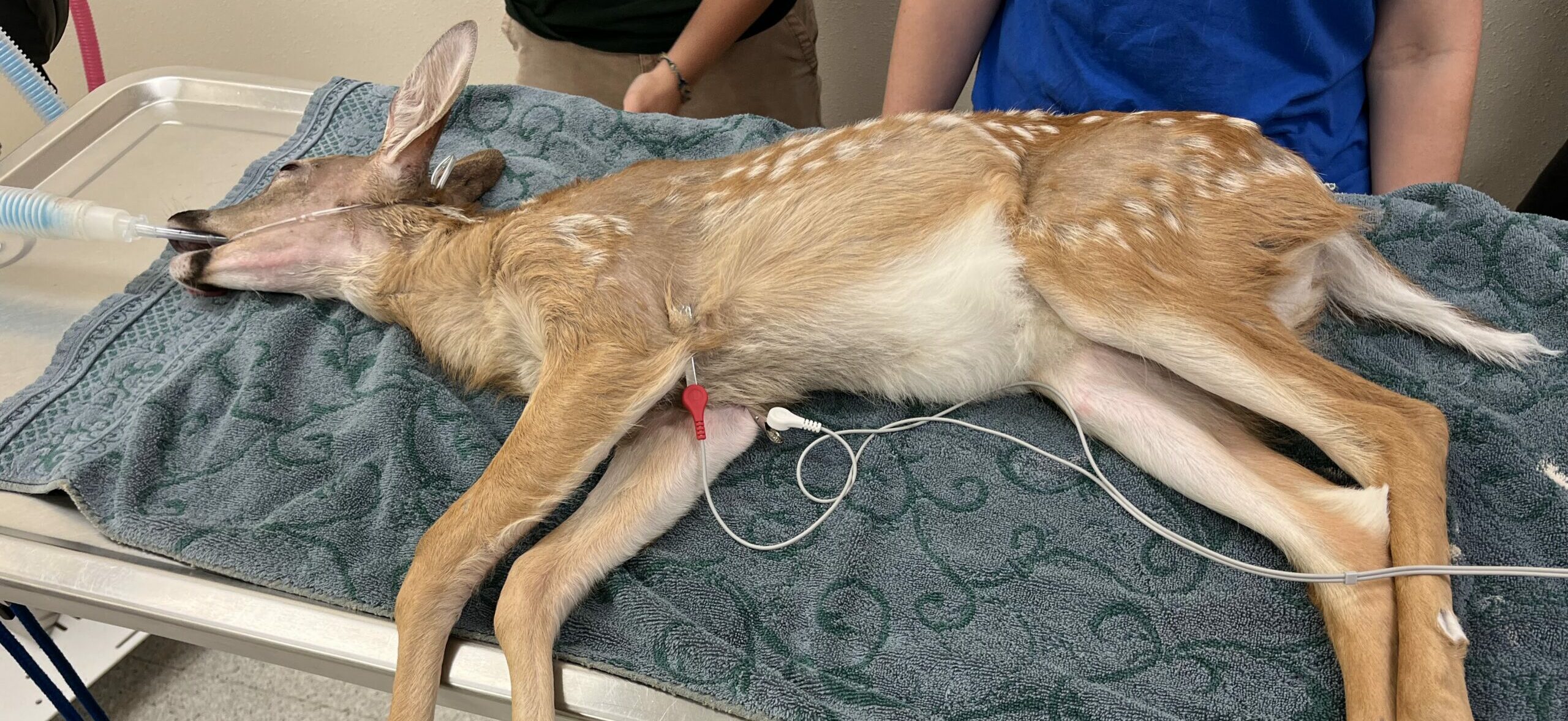
So, after consulting with the veterinarians at the Northwoods Animal Hospital, it was determined that enucleation surgery was the best option to give the fawn a good quality of life. This challenge was undertaken by Dr. David Theurkauff, one of the brilliant and compassionate doctors at the Animal Hospital. The surgery began with the fawn placed under general anesthesia. The hair around his eye and face was carefully clipped and the skin was sterilized with surgical scrub (chlorhexidine). The eyelids of the affected eye were then sutured together before making the incisions to remove the eye. The procedure involved meticulously dissecting the eye from its surrounding tissues, severing the optic nerve, ligating it and also ensuring that any bleeding blood vessels were clamped and tied off to prevent bleeding. Once the eye, along with the eyelids and tear glands, was removed, the skin was sutured over the empty socket with absorbable monocryl sutures, placed in such a way that none of the stitches were visible from the outside.
The surgery was a success! The fawn awoke from anesthesia, now free from the pain that had plagued him for so long. Enucleation surgeries like this one usually have a good success rate and risks of complications are rare, often giving animals not just relief but a new lease on life. In this case, his missing eye posed little threat to his ability to live in the wild. Deer are herd animals; they rely on each other for protection and signal danger to one another, so the loss of one eye wouldn’t significantly impact his chances of survival.
Over the next few weeks, we continued to care for the fawn, treating his other wounds and ensuring he grew strong enough to survive on his own. We usually wait to release fawns until they are old enough to go their separate ways from their mothers in the wild, ensuring they have the best possible chance of survival. When he was finally ready, after a total of 113 days of being in our care, we released him back into the wild along with a few other fawns we were rehabilitating, where they could join a herd and thrive. Releasing him was a bittersweet moment, as it always is.
Enucleation surgery, though often seen as drastic, is a testament to the adaptability and resilience of animals. While it may be challenging for human pet parents to accept when it comes to their pets, the animals themselves often cope remarkably well. Domestic pets like dogs can adapt even if they lose both their eyes, relying on other senses and the care of their owners. Wild animals, however, present a unique challenge. Their survival instincts and natural behaviors, such as hunting or hiding from predators, can be affected. Yet, even in the wild, animals demonstrate an incredible ability to adapt to their circumstances, often way better than humans can. Where a person might struggle with the psychological effects of losing an eye, animals live in the moment, focusing on survival and recovery. This young deer’s story is a powerful example of how animals don’t dwell on their limitations but instead, find new ways to thrive and navigate their environment despite their traumas, relying on their instincts, their peers, and the natural resilience that allows them to overcome adversity. It’s a reminder that resilience is not just a trait, it’s a way of life. With the right care and intervention, even the most vulnerable creatures can be given the opportunity to flourish once more, demonstrating the power of veterinary medicine in giving them a second chance at life.
References:
- https://www.ncbi.nlm.nih.gov/books/NBK562144/#:~:text=Enucleation%20is%20the%20removal%20of,challenging%20therapeutic%20decisions%20to%20make.
- https://www.eyevet.ie/wp-content/uploads/2009/02/enucleation-in-companion-animals-feb-2008-ivj.pdf
- https://www.deerrepellentpacks.com/how-deer-defend-themselves-from-predators
Author: Kiki Raj